


Here we begin the ECG Challenge series, with practical cases to test and sharpen your ECG interpretation.
The idea is simple: analyze the tracing systematically, identify the key findings, and integrate them with the clinical context.
Let’s get to the case.
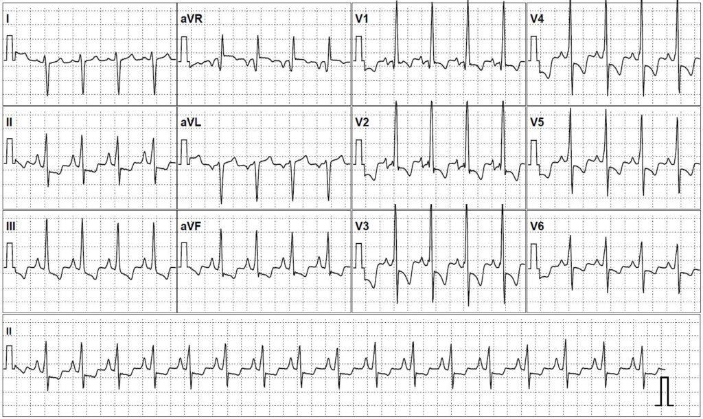
Question
What explains this QRS pattern seen in V1?
Answer and Discussion
1) QRS Morphology in V1
V1 shows a qR pattern, with a QRS duration of around 100 ms.
- Narrow QRS (no right bundle branch block)
- Small initial q wave
- Dominant R wave in V1
This pattern characterizes the Sodi-Pallares sign.
It is an indirect sign of right atrial enlargement, and may also appear as Qr or qRs in V1, in the absence of right bundle branch block.
2) P-Wave Assessment
The P waves are peaked, with an amplitude > 2.5 mm in lead II, consistent with P pulmonale, a classic criterion for right atrial enlargement.
3) QRS Electrical Axis
There is right axis deviation of the QRS, reinforcing the electrical predominance of the right-sided chambers.
4) Signs of Right Ventricular Hypertrophy
In addition, there are tall R waves in V2 and V3, with deep S waves in V6, a combination consistent with right ventricular hypertrophy.
Putting It All Together
- qR pattern in V1 (Sodi-Pallares sign)
- P pulmonale
- Right axis deviation
- Criteria for right ventricular hypertrophy
The tracing is consistent with right heart chamber enlargement. Clinically, the patient is a young adult with severe primary pulmonary hypertension.
Key point: a qR pattern in V1 with a QRS of ~100 ms should not automatically be interpreted as right bundle branch block. QRS duration is decisive in this differentiation.
Learning Points
- A narrow QRS with qR in V1 may indicate the Sodi-Pallares sign.
- Always assess the P wave as well.
- Integrating the whole tracing is worth more than any single criterion.
- Pulmonary hypertension usually produces a right-chamber electrical pattern.
References
- Scheffer MK, De Marchi MFN, de Alencar Neto JN, Felicioni SP. Eletrocardiograma de A a Z. São Paulo: Manole; 2024.
- Surawicz B, Knilans TK. Chou's Electrocardiography in Clinical Practice. 6th ed. Philadelphia: Saunders; 2008.
- Bayés de Luna A, M Fiol-Sala, A Bayés-Genís, A Baranchuk. Clinical Electrocardiography: A Textbook. 5th ed. Hoboken: Wiley; 2021.{index=2}



