


Clinical Case
A 54-year-old previously healthy man is admitted to the emergency department with sudden chest pain and progressive dyspnea for approximately 2 hours.
He denies fever, cough or any cardiac history.
On examination:
- RR: 26 breaths/min
- Oxygen saturation: 90% on room air
- Normal heart sounds, no murmurs
- Breath sounds present, slightly reduced at the right base
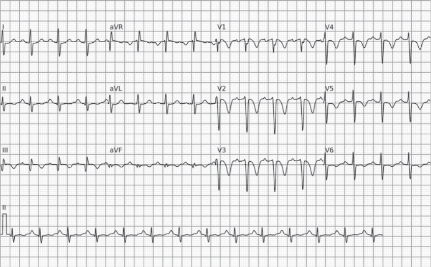
A 12-lead ECG was obtained on admission.
Question
Which electrocardiographic finding is most suggestive of the clinical diagnosis? What is the appropriate initial management?
Answer and Discussion
1) Main ECG finding
The tracing shows:
- Sinus tachycardia
- S1Q3T3 pattern
Characterized by:
- A deep S wave in lead I
- A Q wave in lead III
- An inverted T wave in lead III
This pattern is classically associated with pulmonary embolism (PE).
2) Pathophysiological interpretation
PE causes a sudden increase in pulmonary vascular resistance, leading to:
- Acute right ventricular strain
- Right axis deviation
- Secondary repolarization changes
The S1Q3T3 pattern reflects this acute right ventricular strain, although it has low sensitivity.
3) Other possible findings in PE
- Sinus tachycardia (most frequent finding)
- Right axis deviation
- New or incomplete right bundle branch block
- T-wave inversion in V1–V4
- P pulmonale
- Nonspecific ST-T changes
The ECG may be normal. No single finding confirms the diagnosis.
4) Initial management
When clinical suspicion of PE is high:
- Oxygen therapy
- Hemodynamic risk stratification
- Order a contrast-enhanced CT pulmonary angiogram (if stable)
- Early initiation of anticoagulation, if there are no contraindications
In unstable patients, consider immediate evaluation for systemic thrombolysis.
Practical Points
- Sinus tachycardia is the most common finding in PE.
- S1Q3T3 is classic but has low sensitivity.
- The ECG helps raise suspicion but does not confirm the diagnosis.
- The treatment decision depends on hemodynamic risk.



