


We continue the ECG Challenge series. In this case, the goal is to answer two fundamental questions in a patient with STEMI:
- Which wall is affected?
- What is the culprit artery?
Clinical Case
Patient with typical chest pain and an ECG consistent with STEMI.
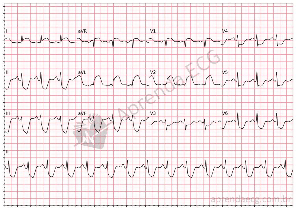
Question
Which wall is affected and what is the culprit artery?
Answer and Discussion
1) Distribution of ST elevation
The tracing shows:
- ST elevation in I and aVL
- ST elevation in V2
- ST depression in the inferior leads
This combination points the reasoning toward involvement of the mid-anterior wall.
2) Topographic correlation according to Bayés de Luna
According to the topographic classification proposed by Bayés de Luna, based on electrocardiographic correlation with cardiac magnetic resonance imaging, leads I, aVL and V2 correspond to the mid-anterior wall.
This region represents an intermediate territory of the anterior wall of the left ventricle, corresponding neither to the classic extensive anterior infarction (V1–V4) nor to isolated lateral involvement.
When there is ST elevation in this specific set of leads, the artery most frequently involved is the first diagonal branch (D1) of the left anterior descending artery.
Anatomically, the first diagonal supplies precisely this anteromedial portion of the left ventricle, which explains the distribution of ST elevation seen on the ECG.
3) Diagnostic integration
Conclusion:
- Affected wall: mid-anterior
- Culprit artery: first diagonal branch (D1) of the left anterior descending artery
The pattern may be subtler than in extensive anterior STEMI due to proximal LAD occlusion, which increases the risk of underdiagnosis.
However, this is an acute coronary occlusion and requires an immediate reperfusion strategy.
Key Points for Practice
- I + aVL + V2 correspond to the mid-anterior wall.
- This pattern suggests occlusion of the first diagonal branch.
- Not every anterior STEMI involves the proximal LAD.
- Modern topographic classification is based on correlation with magnetic resonance imaging.
References
- Durant E, Singh A. Acute first diagonal artery occlusion: a characteristic pattern of ST elevation . Am J Emerg Med. 2015;33:1326.e3-5.
- Littmann L. South African flag sign: a teaching tool for easier ECG recognition . Am J Emerg Med. 2016;34(1):107-109.
- Rajendran G, Mahalingam S, Kagne R, Nathan B. The South African flag sign – an electrocardiographic sign to predict the culprit artery . QJM. 2021;114(9):665-666.
- Monaghan M, Sreenivasa S. A Red Flag ECG . Circulation. 2020;142:1871–1874.



