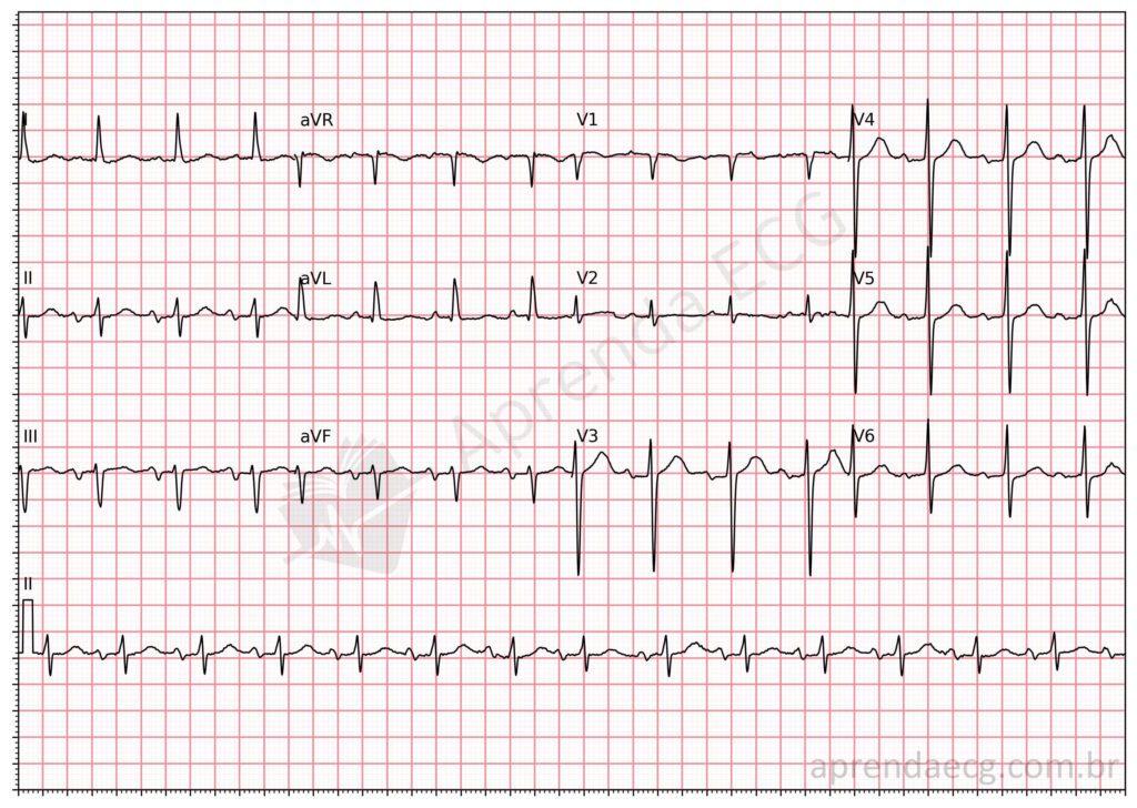
The P wave in leads II, III and aVF should last up to 120 ms. When it exceeds this limit, it is considered wide and may suggest atrial abnormalities. However, there is an interesting debate involving old myths and paradigms about the true meaning of this finding: is it related to left atrial enlargement (LAE) or, in fact, to interatrial block (IAB)?
Deconstructing the paradigm
To answer the question posed in this article, it is important to revisit some concepts of evidence-based medicine.
A P wave lasting 120 ms or more in leads II, III or aVF, when used as a criterion for reporting LAE, has a positive likelihood ratio (LR+) of 0.94 and a negative likelihood ratio (LR-) of 1.14. These values indicate that the presence of this finding lowers the probability of LAE (because the LR+ is below 1), while its absence raises it (because the LR- is above 1). Based on these data, it is clear that P-wave duration is not a reliable criterion for diagnosing LAE, and the Morris index is a more accurate criterion for this purpose.
On the other hand, studies using echocardiography and magnetic resonance imaging show that a widened P wave is more closely associated with IAB than with LAE. Thus, a P wave lasting 120 ms or more in II, III and aVF indicates IAB, not LAE.
This shows that interpretation of a widened P wave follows a logic similar to that of the QRS complex in the ventricles: while P-wave widening indicates IAB, a tall P wave may suggest atrial enlargement, just as a widened QRS points to bundle branch block and a tall QRS to ventricular hypertrophy.
Learn more
To review the main concepts covered here, see our posts on Left Atrial Enlargement and Interatrial Block.
References
- Scheffer MK, De Marchi MFN, de Alencar Neto JN, Felicioni SP. Eletrocardiograma de A a Z. São Paulo: Manole, 2024.
- Alencar JN. Tratado de ECG. São Paulo: Manole, 2022.
- Tsao CW, Josephson ME, Hauser TH, O’Halloran TD, Agarwal A, Manning WJ, et al. Accuracy of electrocardiographic criteria for atrial enlargement: validation with cardiovascular magnetic resonance. J Cardiovasc Magn Reson. 2008;10(1):1-7.