Interatrial block (IAB) is a fascinating and often underestimated topic in electrocardiogram (ECG) study. Although frequently confused with left atrial enlargement (LAE), these blocks have unique features and important clinical implications that every cardiologist should know. In this article, we take an in-depth look at what IABs are, how to identify them and why they are so relevant to clinical practice.
What Is Interatrial Block?
IAB occurs when there is a delay or block in electrical conduction between the atria, usually through Bachmann's bundle, a structure that carries the electrical impulse generated in the sinus node of the right atrium to the left atrium. On the ECG, this delay appears as increased P-wave duration and changes in its morphology.
Left Atrial Enlargement and Increased P-Wave Duration
Increased P-wave duration can be mistaken for LAE but must be assessed carefully. A prolonged P wave occurs in the presence of IAB and should not automatically be interpreted as LAE. Studies show that a prolonged P wave has a positive likelihood ratio (LR+) of 0.94 and a negative likelihood ratio (LR-) of 1.14 for the diagnosis of LAE, indicating that on its own it may not be useful for this diagnosis. For the electrocardiographic diagnosis of LAE, we recommend using the Morris index.
Pathophysiological Mechanism
Interatrial conduction normally occurs through Bachmann's bundle. When this bundle is delayed or blocked, the electrical impulse may be diverted to other, less efficient routes, such as the interatrial septum or the coronary sinus, producing the patterns seen in IAB. This process may be triggered by atrial remodeling, fibrosis or other structural changes that affect electrical conduction.
Classification of Interatrial Block
IABs are classified into three degrees:
- First-degree (partial) IAB: characterized by a P wave lasting 120 ms or more, usually with a biphasic morphology in leads such as II, III and aVF. Conduction is delayed but not completely blocked.
- Second-degree IAB: interatrial conduction may be normal or show features of first- or third-degree IAB intermittently on the same ECG or at different times. This is known as “atrial aberrancy.”
- Third-degree (advanced) IAB: the impulse is blocked in the region of Bachmann's bundle and the fossa ovalis, allowing conduction only through the coronary sinus. The P wave is prolonged and shows a plus-minus morphology in the inferior leads.
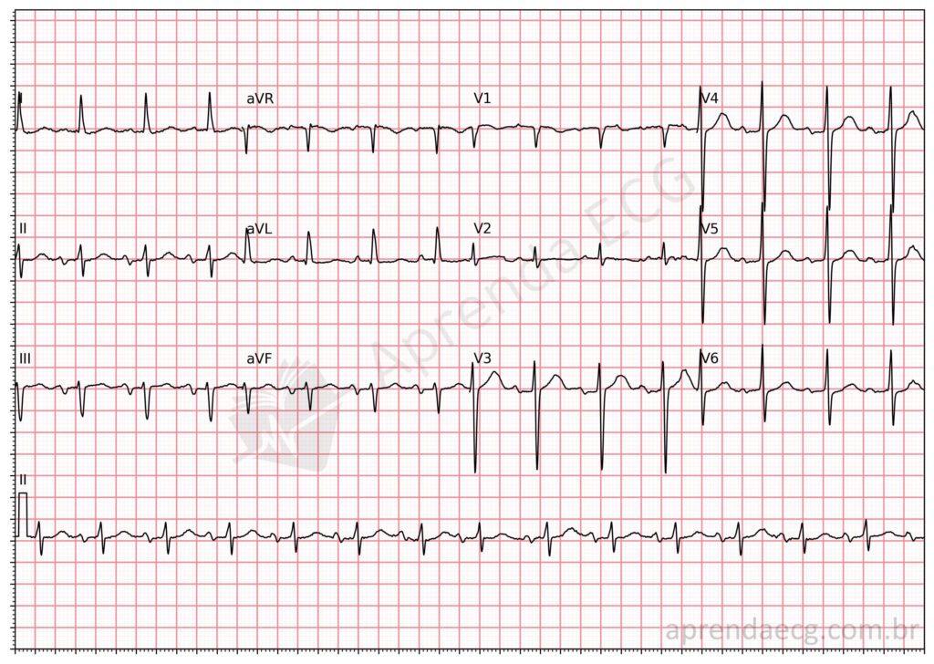
Explore our ECG library for more examples of ECGs with IAB.
Clinical and Prognostic Implications
IAB, especially the advanced form, is associated with an increased risk of supraventricular arrhythmias such as atrial fibrillation (AF). The BAYES Registry, published in 2020, showed that advanced IAB is independently associated with ischemic cerebral events, underscoring the importance of identifying and appropriately monitoring these patients.
Atypical Interatrial Block
Atypical patterns of advanced IAB (A-IAB) have recently been identified, characterized by variations in P-wave morphology or duration. These atypical patterns are not yet fully understood, but they may represent an important subgroup of patients at high risk of AF and stroke. The true prevalence of these patterns in different clinical settings, as well as their prognostic value, remains to be determined.
Conclusion
IABs are important disorders affecting electrical conduction between the atria. Their classification into degrees and an understanding of the pathophysiological mechanisms are essential to distinguish IAB from LAE. In addition, their clinical and prognostic implications highlight the importance of careful evaluation and monitoring of affected patients.
References
- Bayés De Luna A, Platonov P, Cosio FG, Cygankiewicz I, Pastore C, Baranowski R, et al. Interatrial blocks. A separate entity from left atrial enlargement: A consensus report. J Electrocardiol [Internet]. 2012;45(5):445–51.
- Martínez-Sellés M, Elosua R, Ibarrola M, De Andrés M, Díez-Villanueva P, Bayés-Genis A, et al. Advanced interatrial block and P-wave duration are associated with atrial fibrillation and stroke in older adults with heart disease: The BAYES registry. Europace. 2020;22(7):1001–8.
- Bayés de Luna A, Escobar-Robledo LA, Aristizabal D, Weir Restrepo D, Mendieta G, Massó van Roessel A, et al. Atypical advanced interatrial blocks: Definition and electrocardiographic recognition. J Electrocardiol. 2018;51(6):1091–3.
- Tsao CW, Josephson ME, Hauser TH, O’Halloran TD, et al. Accuracy of electrocardiographic criteria for atrial enlargement: Validation with cardiovascular magnetic resonance. J Cardiovasc Magn Reson. 2008;10(1):1–7.