

Interatrial block (IAB) refers to delayed conduction of the impulse from the right atrium to the left atrium through Bachmann's bundle. It can be first degree (P-wave duration > 120 ms), third degree (prolonged, biphasic [±] P wave in the inferior leads), or second degree when these patterns appear transiently in the same lead (atrial aberrancy).
Electrocardiographic Criteria
- Partial or first-degree IAB: P wave ≥ 120 ms in leads II, III, and aVF
- Second-degree IAB: intermittent alternation between normal interatrial conduction and the first- or third-degree pattern
- Advanced or third-degree IAB: P wave ≥ 120 ms in leads II, III, and aVF with plus-minus morphology
Clinical Implications of Interatrial Block
First-degree interatrial blocks are very common, especially in older adults, and are associated with atrial fibrillation and increased all-cause and cardiovascular mortality. Third-degree interatrial blocks are less frequent, but they are strong markers of left atrial enlargement (LAE) and paroxysmal supraventricular tachycardias (Bayés syndrome).
ECG in Interatrial Block
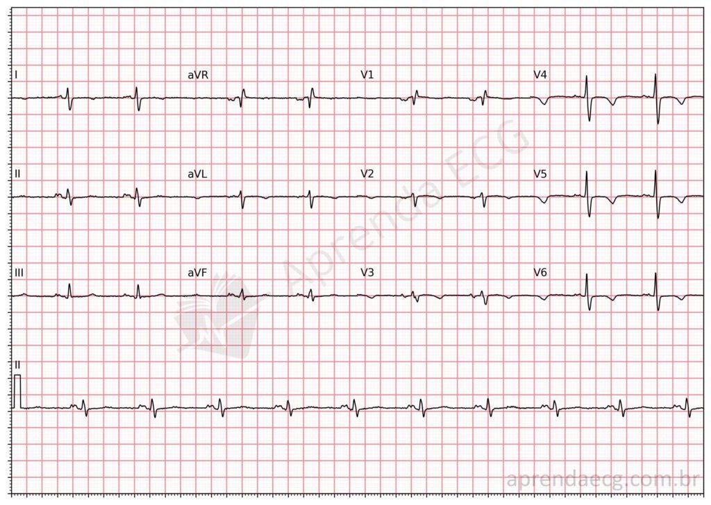
Partial interatrial block. Delayed conduction from the right atrium to the left atrium through Bachmann's bundle increases P-wave duration in the inferior leads (II, III, and aVF) and causes notching of this wave.
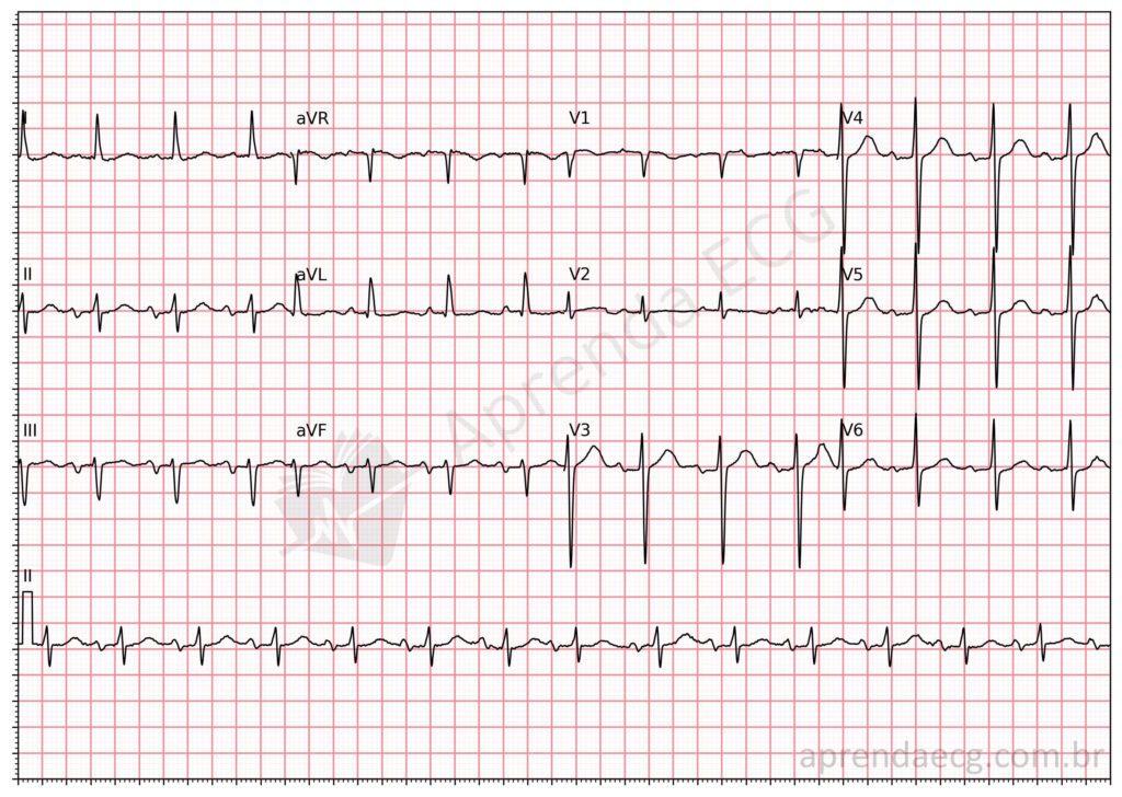
Advanced interatrial block. In advanced interatrial block, the electrical impulse is blocked in the region of Bachmann's bundle, so left atrial activation occurs through the coronary sinus, that is, from bottom to top, producing a P wave longer than 120 ms in the inferior leads with plus-minus morphology.
Further Reading
Related Topics
References
- Bayés De Luna A, Platonov P, Cosio FG, Cygankiewicz I, et al. Interatrial blocks. A separate entity from left atrial enlargement: A consensus report. J Electrocardiol. 2012;45(5):445–51.


