

Right atrial enlargement (RAE) is an electrocardiographic abnormality characterized by changes in P-wave morphology that indicate increased right atrial pressure or volume. On the ECG, it usually manifests as a higher-amplitude P wave in the inferior leads (especially lead II) and in V1, where the P wave may have a more peaked morphology.
ECG Criteria
- P wave > 2.5 mm in II, III, and aVF
- Positive portion of the P wave > 1.5 mm in V1 and V2
- Sodi-Pallares sign: QR or qR complexes in V1
- Peñaloza-Tranchesi-Reeves sign: QRS complex < 4 mm in V1, with an increase in amplitude greater than 5-fold from V1 to V2
ECG in Right Atrial Enlargement
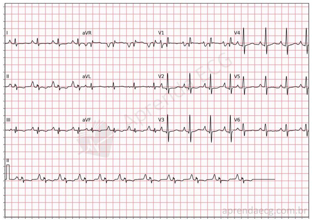
Biatrial enlargement. Right atrial enlargement can be diagnosed by a P wave with amplitude greater than 2.5 mm in the inferior wall (II, III, and aVF) and by the Sodi-Pallares sign (qR complex in V1). In addition, this ECG shows the Morris index in V1, indicating left atrial enlargement. This is therefore biatrial enlargement.
Related Topics
References
- Samesima N, God EG, Kruse JCL, Leal MG, et al. Diretriz da Sociedade Brasileira de Cardiologia sobre a Análise e Emissão de Laudos Eletrocardiográficos – 2022. Arq Bras Cardiol. 2022;93(3 Suppl 2):2–19.
- Sodi-Pallares D, Bisteini A, Herrmanndg GR. Some views on the significance of qR and QR type complexes in right precordial leads in the absence of myocardial infarction. Am Heart J. 1952;43(5):716–34.
- Reeves WC, Hallahan W, Schwiter EJ, Ciotola TJ, et al. Two-dimensional echocardiographic assessment of electrocardiographic criteria for right atrial enlargement. Circulation. 1981;64(2):387–91.


