

Left atrial enlargement is an ECG abnormality that indicates increased pressure or volume in the left atrium, usually related to conditions that overload this cardiac chamber. On the electrocardiographic tracing, it typically manifests as a P wave with a deeper and longer negative terminal portion in V1.
ECG Criteria
- Morris index: increased negative component (slow, deep terminal portion) in lead V1 (≥ 40 ms in duration and ≥ 1 mm in amplitude)
Increased P-Wave Duration > 120 ms
A P wave lasting more than 120 ms in leads II, III, or aVF, when not accompanied by the Morris index, usually does not indicate left atrial enlargement (LAE). Instead, this P-wave morphology is more closely associated with interatrial block, a conduction disorder between the atria that delays the electrical impulse through Bachmann's bundle, which is responsible for activating the left atrium.
ECG in Left Atrial Enlargement
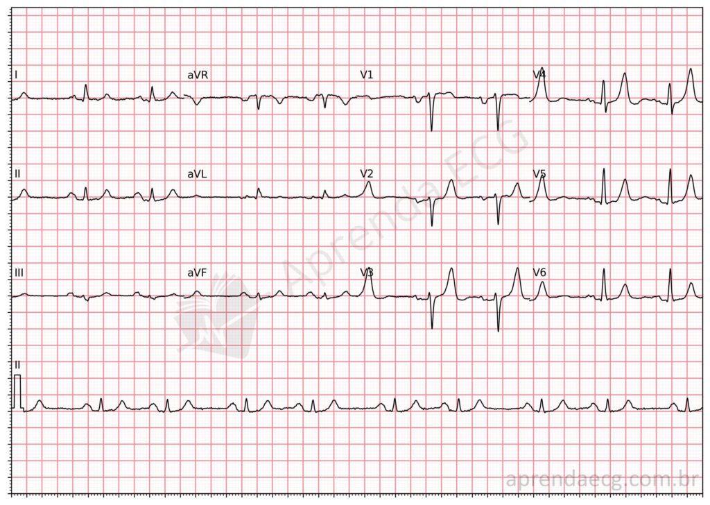
Left atrial enlargement. For the electrocardiographic diagnosis of LAE, we use the Morris index, that is, the negative portion of the P wave in V1 is longer than 40 ms and deeper than 1 mm.
Further Reading
Related Topics
References
- Alencar Neto JN de. Tratado de ECG. Salvador: Sanar; 2022. 816 p.
- Bayés De Luna A, Platonov P, Cosio FG, Cygankiewicz I, et al. Interatrial blocks. A separate entity from left atrial enlargement: A consensus report. J Electrocardiol. 2012;45(5):445–51.


