

Left ventricular hypertrophy (LVH) is suggested on the ECG by changes in the QRS complexes, with increased R- and S-wave voltage, mainly in the leads that reflect left ventricular activity. Criteria such as Sokolow-Lyon, Cornell, and Peguero-Lo Presti help identify these signs on the ECG and support the diagnosis. Early detection of LVH is important, as it is associated with a higher risk of cardiovascular events.
ECG Criteria
The ECG criteria for LVH are based on QRS complex voltage. However, many such criteria exist in the literature. In our opinion, the most important are:
- Peguero-Lo Presti: deepest S wave + S wave in V4 > 23 mm in women and > 28 mm in men
- Cornell: S wave in V3 + R wave in aVL > 20 mm in women and > 28 mm in men
- Sokolow-Lyon: S wave in V1 + R wave in V5 or V6 > 35 mm
- R in aVL: R wave in aVL > 11 mm
- Gubner-Ungerleider: R wave in I + S wave in III > 25 mm
Point Score System
Another way to diagnose LVH is the Romhilt-Estes point score. The diagnosis is confirmed with 5 or more points and is probable with 4 points.
- Increased amplitude: R or S wave > 20 mm in the frontal plane or > 30 mm in the horizontal plane (3 points)
- ST-T changes (strain) without digitalis (3 points)
- LAE: Morris index (3 points)
- QRS axis deviation beyond -30° (2 points)
- QRS ≥ 90 ms without a bundle branch block pattern (1 point)
- Ventricular activation time ≥ 50 ms in V5 and V6 (1 point)
- ST-T changes (strain) with digitalis (1 point)
Other ECG Changes in LVH
- Secondary ventricular repolarization abnormalities with ST-segment depression and negative, asymmetric T waves in the lateral leads (I, aVL, V5, and V6): strain
- Slight increase in QRS duration
- Leftward deviation of the QRS axis
- Mild QT interval prolongation
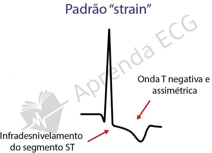
ECG in Left Ventricular Hypertrophy
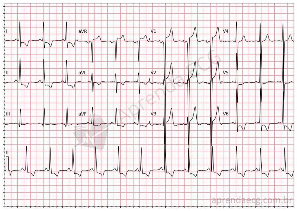
Left ventricular hypertrophy. Male patient whose ECG meets electrocardiographic criteria for LVH: Sokolow-Lyon and Peguero-Lo Presti. There are also ventricular repolarization abnormalities with a strain pattern.
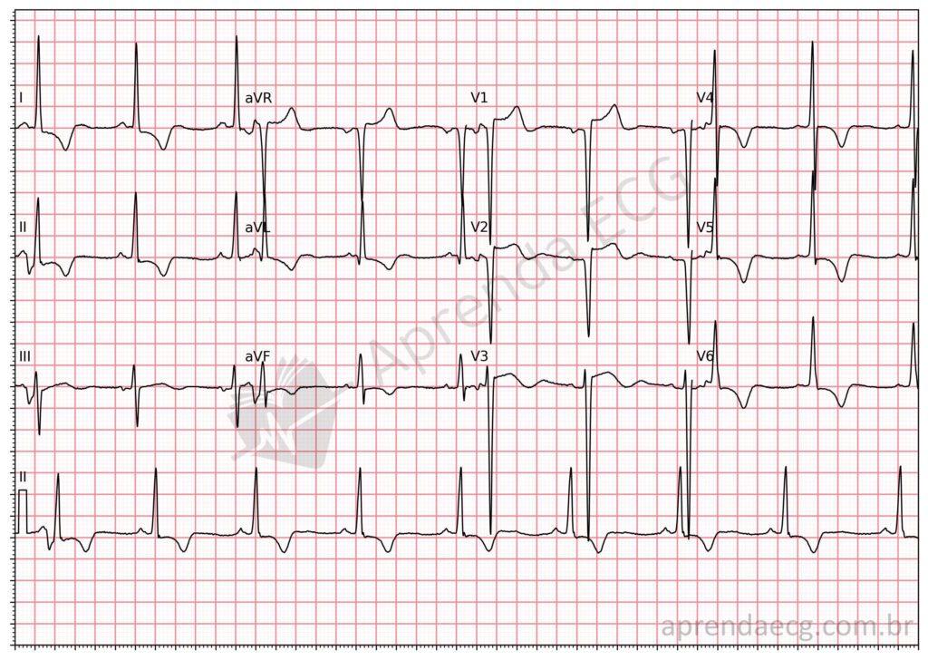
Left ventricular hypertrophy. Male patient whose ECG meets the Sokolow-Lyon, Peguero-Lo Presti, and R in aVL criteria. There is also a strain-type ventricular repolarization abnormality.
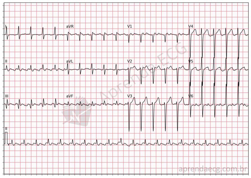
Left ventricular hypertrophy. In this example, the underlying rhythm is atrial flutter, and LVH can be diagnosed by the Peguero-Lo Presti criterion (deepest S wave on the ECG plus the S wave in V4).
Further Reading
Related Topics
References
- Scheffer MK, De Marchi MFN, de Alencar Neto JN, Felicioni SP. Eletrocardiograma de A a Z. São Paulo: Manole, 2024.
- Hancock EW, Deal BJ, Mirvis DM, Okin P, et al. AHA/ACCF/HRS Recommendations for the Standardization and Interpretation of the Electrocardiogram. Part V: Electrocardiogram Changes Associated With Cardiac Chamber Hypertrophy A Scientific Statement From the American Heart Association Electrocardiography. J Am Coll Cardiol. 2009;53(11):992–1002.
- Peguero JG, Lo Presti S, Perez J, Issa O, et al. Electrocardiographic Criteria for the Diagnosis of Left Ventricular Hypertrophy. J Am Coll Cardiol. 2017;69(13):1694–703.
- Romhilt DW, Estes EH. A point-score system for the ECG diagnosis of left ventricular hypertrophy. Am Heart J. 1968;75(6):752–8.


