

Biventricular hypertrophy occurs when both the left and the right ventricles are enlarged, and it shows up on the ECG as combined signs of right and left ventricular hypertrophy. The tracing may show features such as increased R waves in V1 (indicating right ventricular hypertrophy) and in the left-sided leads (indicating left ventricular hypertrophy), as well as electrical axis deviation and QRS complex abnormalities.
ECG Criteria
- Frontal plane QRS axis deviated to the right, associated with voltage criteria for LVH
- ECG typical of RVH, associated with one or more of the following:
- Deep Q waves in V5 and V6 and in the inferior leads;
- Increased R-wave voltage in V5 and V6;
- S in V1 and V2 + R in V5 and V6 meeting the Sokolow criterion;
- Ventricular activation time in V6 of 40 ms or more;
- Large isodiphasic QRS complexes, R/S type > 50 mm, in the mid-precordial leads V2 to V4 (Katz-Wachtel phenomenon)
ECG in Biventricular Hypertrophy
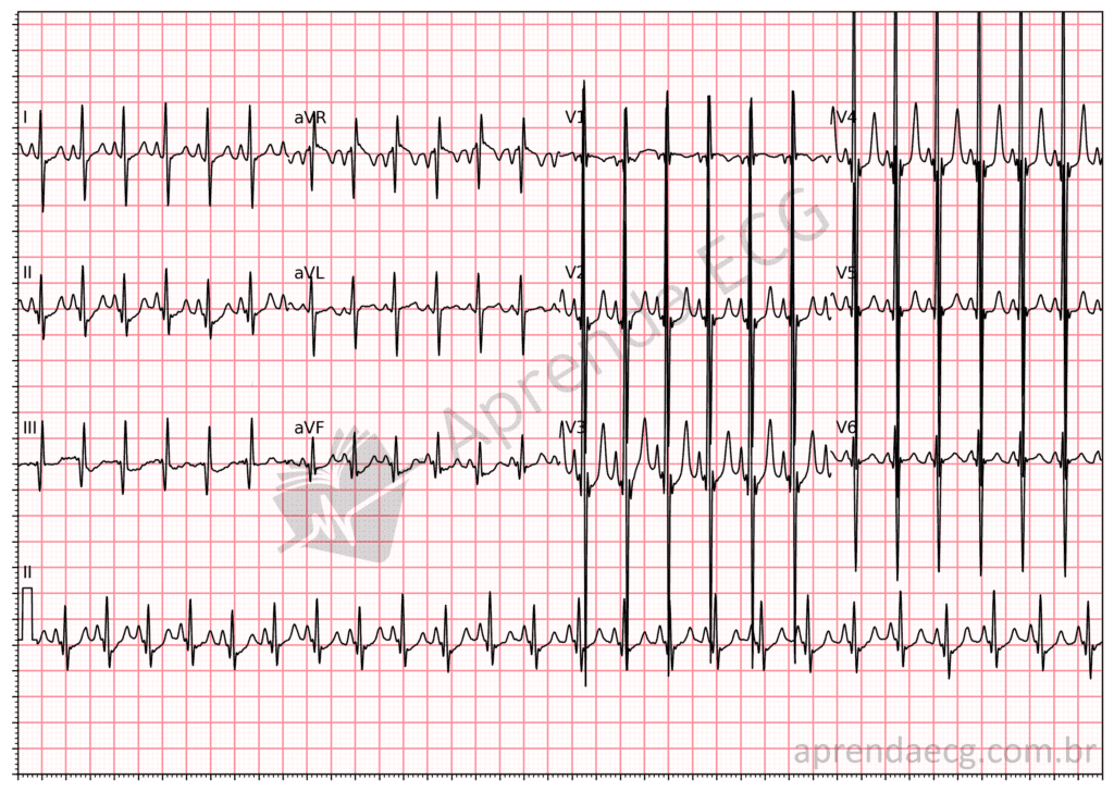
Biventricular hypertrophy. ECG showing the Katz-Wachtel phenomenon in a 6-month-old child with congenital heart disease.
Related Topics
References
- Samesima N, God EG, Kruse JCL, Leal MG, et al. Diretriz da Sociedade Brasileira de Cardiologia sobre a Análise e Emissão de Laudos Eletrocardiográficos – 2022. Arq Bras Cardiol. 2022;93(3 Suppl 2):2–19


