
What Can QRS Fragmentation Reveal?
The electrocardiogram (ECG) is a powerful tool capable of identifying areas of myocardial fibrosis and cardiac scarring. You are probably familiar with the classic Q waves that develop in the course of an acute myocardial infarction with coronary occlusion. But what if we told you there is another marker, less well known but equally important?
QRS fragmentation (fQRS), a more recently described pattern, reveals subtle abnormalities in electrical conduction that may indicate myocardial fibrosis, predict adverse cardiac events and even signal a higher risk of mortality. In this article, we explore the criteria, mechanisms and clinical implications of this essential marker for modern electrocardiographic practice.
Pathological Q Waves: How and When Do They Form?
Pathological Q waves appear when an area of myocardium undergoes transmural infarction, resulting in the absence of electrical activity from that region. This is seen at the beginning of the QRS because the affected area is depolarized very early, in the first 40 to 50 ms of the ventricular electrical cycle. This creates an electrical force directed away from the infarcted region, producing a Q wave in the corresponding leads.
However, whether a pathological Q wave develops depends on the size and location of the infarction. Small scars or deeper areas may not produce visible Q waves on the ECG, limiting their sensitivity as a marker of myocardial scar.
QRS Fragmentation: Definition and Criteria
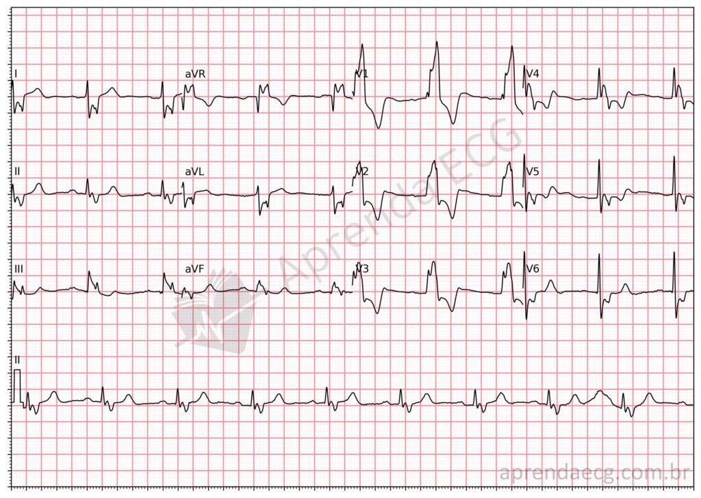
QRS fragmentation, on the other hand, reflects areas of scar or myocardial fibrosis that disrupt electrical conduction in the middle or at the end of the QRS, unlike Q waves. These regions depolarize late because of altered intramyocardial conduction caused by fibrous tissue or structural irregularities. The result is additional waves, such as notches or late R’ waves, that fragment the QRS complex. These notches or additional waves are seen in two or more contiguous leads corresponding to coronary territories. The fragmentation pattern can be found in both narrow QRS (<120 ms) and wide QRS, as in bundle branch block or paced rhythm.
ECG Diagnostic Criteria
- An additional R’ wave, a notch in the nadir of the S wave or fragmentation with ≥ 1 R’ in at least 2 contiguous leads
- In wide QRS (≥120 ms), fragmentation is defined by ≥ 2 notches in the R or S wave, also in contiguous leads
Why Does It Happen?
- Myocardial scars: interrupt normal electrical propagation, causing slow conduction and local block.
- Fibrous tissue: creates irregular conduction pathways, leading to delayed electrical activation in specific areas of the myocardium.
- Focus of late depolarization: while Q waves indicate absent depolarization in an infarcted region, fQRS signals areas with delayed conduction within the cycle.
Etiologies
The most common etiologies include prior myocardial infarction, in which areas of necrosis evolve into fibrotic scars, and coronary artery disease with chronic ischemia. Other causes include cardiomyopathies, such as dilated and hypertrophic cardiomyopathy, which promote remodeling and ventricular dysfunction, as well as infiltrative diseases such as sarcoidosis and amyloidosis, which affect electrical conduction by altering myocardial architecture. It can also be seen in non-ischemic conditions such as myocarditis, and even in some systemic diseases such as obstructive sleep apnea and autoimmune diseases, which may secondarily impair cardiac conduction.
Comparison of Q Waves and fQRS
| Feature | Q wave | QRS fragmentation (fQRS) |
|---|---|---|
| Timing | Beginning of the QRS (first 40-50 ms) | Middle and end of the QRS |
| Main cause | Absent initial depolarization | Delayed depolarization |
| Indicative of | Transmural infarction | Scar or abnormal conduction |
| Sensitivity | Lower (36%-51%) | Higher (68%-86%) |
| Specificity | High (97%) | Moderate (80%-92%) |
Clinical and Prognostic Implications
Detection of Myocardial Scars
fQRS is a reliable marker for locating scars in specific territories, even when Q waves are absent. For example:
- Anterior: leads V1 to V5.
- Lateral: leads I, aVL, V6.
- Inferior: leads II, III, aVF.
Cardiovascular Prognosis
Studies show that fQRS is associated with:
- Higher risk of all-cause and cardiovascular mortality.
- Prediction of events such as heart failure and ventricular arrhythmias.
- Worse in-hospital outcomes in patients with acute coronary syndromes.
Key Points
- The Q wave reflects absent depolarization in infarcted areas, visible in the first 40-50 ms of the QRS.
- QRS fragmentation arises from late depolarization, affecting the middle and end of the QRS.
- fQRS is more sensitive than Q waves for detecting myocardial scars and also offers significant prognostic value.
- Identifying fQRS on the ECG is a simple but essential step in clinical practice.
References
- Das MK, Khan B, Jacob S, Kumar A, Mahenthiran J. Significance of a fragmented QRS complex versus a Q wave in patients with coronary artery disease. Circulation. 2006;113(21):2495–501.
- Das MK, Suradi H, Maskoun W, Michael MA, Shen C, Peng J, et al. Fragmented wide QRS on a 12-lead ECG: a sign of myocardial scar and poor prognosis. Circ Arrhythm Electrophysiol. 2008;1(4):258–68.
- Das MK, Zipes DP. Fragmented QRS: A predictor of mortality and sudden cardiac death. Hear Rhythm [Internet]. 2009;6(3 SUPPL.):S8–14. Available from: http://dx.doi.org/10.1016/j.hrthm.2008.10.019
- Sadeghi R, Dabbagh VR, Tayyebi M, Zakavi SR, Ayati N. Diagnostic value of fragmented QRS complex in myocardial scar detection: Systematic review and meta-analysis of the literature. Kardiol Pol. 2016;74(4):331–7.