
Introduction
Lateral wall infarction is one of the most challenging types of infarction for healthcare professionals, especially when the occlusion occurs in the left circumflex artery (LCx). It is not always evident on the electrocardiogram (ECG), and the absence of ST-segment elevation can often lead to underdiagnosis. So how can we identify the subtle signs and ensure an accurate diagnosis? Keep reading to discover the patterns that can make all the difference.
Circumflex Artery Occlusion and the ECG
The left circumflex artery (LCx) is one of the main arteries supplying blood to the lateral wall of the left ventricle. The LCx arises from the left main coronary artery, which bifurcates into the left anterior descending artery (LAD) and the circumflex. It runs along the posterior portion of the left atrioventricular groove, giving off branches that mainly supply the lateral wall. When the LCx is occluded, we do not always see ST-segment elevation, which is the classic criterion for infarction. Studies show that LCx occlusion may present with no ST-segment deviation in up to 38% of cases and with isolated ST depression in 15% of cases, while only half of patients show ST elevation during the acute phase.
Electrocardiographic Patterns in LCx Occlusion
The ECG of patients with LCx occlusion may show several patterns beyond ST-segment elevation. These include:
- ST elevation in the lateral leads (I, aVL, V5, V6) or inferolateral leads (I, aVL, V5, V6, II, III and aVF). This pattern may indicate acute coronary occlusion in territories supplied by the circumflex artery.
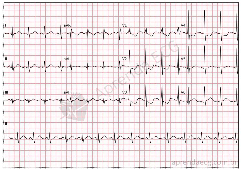
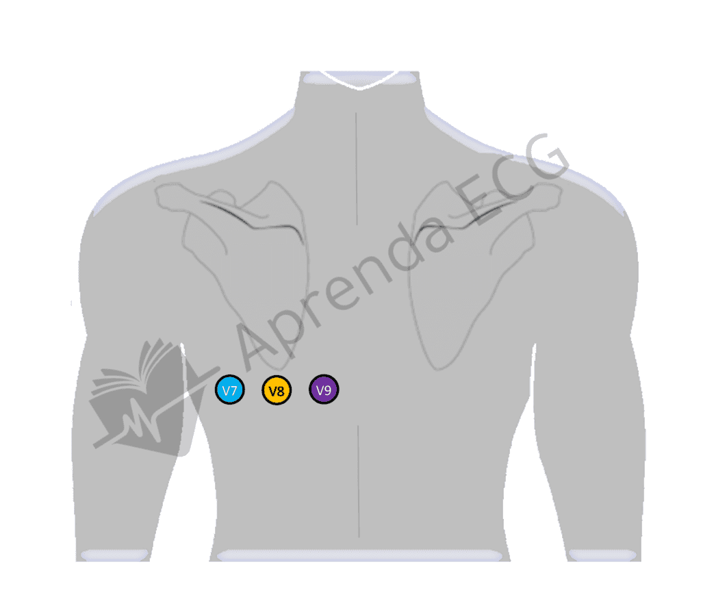
- ST depression in leads V1 to V3, especially when the occlusion is proximal. In this context, it is mandatory to record additional leads, such as V7, V8 and V9, to assess for ST elevation in these leads. About 10 to 15% of acute myocardial infarctions present with isolated ST depression in V1 to V3, frequently associated with LCx occlusion.
- N wave, defined as a deflection or notch at the end of the QRS complex, was observed in 77% of patients with LCx occlusion, with a specificity of 89% for identifying this artery as the culprit.
The Challenge of Diagnosing LCx Occlusion
The frequent absence of ST elevation in patients with LCx occlusion is due to the anatomy and physiology of the artery and to the contribution of collateral circulation. Patients with occlusion proximal to the obtuse marginal branches are at greater risk of showing no obvious ECG changes, which can delay treatment and increase morbidity.
The Importance of Lateral Chest Leads (V7, V8 and V9)
Using the lateral leads (V7-V9) is essential when there is ST depression in leads V1-V3. These additional leads increase the sensitivity of the ECG for detecting LCx occlusion, helping to identify lateral infarctions that could go unnoticed on the standard 12-lead ECG.
ST Elevation in Acute Coronary Occlusion
Our team conducted a systematic review and meta-analysis on the accuracy of ST-segment elevation in the diagnosis of acute coronary occlusion (OMI). The study analyzed the sensitivity and specificity of ST elevation, highlighting the limitations of the current STEMI-NSTEMI paradigm. The results showed that the sensitivity of ST elevation for detecting OMI was only 43.6%, while the specificity was 96.5%. This reveals a significant diagnostic gap, suggesting that more than half of OMI cases may not show classic ST elevation. To learn more about this study, read the full article available here on our site.
Key Points
- Occlusion of the circumflex artery does not always cause ST elevation on the ECG.
- Patterns such as ST depression in V1 to V3 and the presence of an N wave are important for identifying lateral wall infarction.
- Recording posterior chest leads in suspected cases can increase diagnostic accuracy and help detect infarctions that are not evident.
References
- Scheffer MK, De Marchi MFN, de Alencar Neto JN, Felicioni SP. Eletrocardiograma de A a Z. São Paulo: Manole, 2024.
- Geffin R, Triska J, Najjar S, Berman J, Cruse M, Birnbaum Y. Why do we keep missing left circumflex artery myocardial infarctions? J Electrocardiol. 2024;83(December 2023):4–11.
- Birnbaum Y, Bayés de Luna A, Fiol M, Nikus K, Macfarlane P, Gorgels A, et al. Common pitfalls in the interpretation of electrocardiograms from patients with acute coronary syndromes with narrow QRS: a consensus report. J Electrocardiol. 2012 Sep;45(5):463–75.
- Niu T, Fu P, Jia C, Dong Y, Liang C, Cao Q, et al. The delayed activation wave in non-ST-elevation myocardial infarction. Int J Cardiol. 2013;162(2):107–11
- de Alencar Neto JN, Scheffer MK, Correia BP, Franchini KG, Felicioni SP, De Marchi MFN. Systematic review and meta-analysis of diagnostic test accuracy of ST-segment elevation for acute coronary occlusion. Int J Cardiol. 2024 Feb;131889.